Post-Harvest Fungal Diseases of Stored Sweet Potatoes (Ipomoea batatas (L.) Lam) in Three Markets of Jos, Plateau State
-
Nnebechukwu, Ijeoma Adaku

Department of Plant Science and Biotechnology, Faculty of Natural Sciences, University of Jos, Plateau State, Nigeria
Musa Stephanie YoroDepartment of Plant Science and Biotechnology, Faculty of Natural Sciences, University of Jos, Plateau State, Nigeria
P.O. NwadiaroDepartment of Plant Science and Biotechnology, Faculty of Natural Sciences, University of Jos, Plateau State, Nigeria
| Received 16 Feb, 2025 |
Accepted 18 Mar, 2025 |
Published 19 Mar, 2025 |
Background and Objective: One of the greatest problems of food production in Nigeria is that of storage, which leads to post-harvest losses, especially in tubers like sweet potato, which are caused by fungi. This study aimed to isolate, identify, and evaluate the pathogenicity of fungal species associated with sweet potato tuber rot in three markets within Jos, Nigeria. Materials and Methods: Diseased tubers were collected, and fungal isolates were cultured on potato dextrose agar (PDA) which was prepared by boiling 200 g of peeled potatoes in 1 L of distilled water, filtering the infusion, and adding dextrose and agar before sterilization. Fungal isolation was done, where the diseased sweet potato tissues were aseptically excised and inoculated on PDA. Plates were incubated at 25°C for 3-7 days, and fungal colonies were purified through subculturing with streptomycin added to suppress bacterial growth. Identification of fungal isolates was based on morphological characteristics and compared with existing descriptions. Pathogenicity tests involved inoculating healthy, surface-sterilized sweet potato tubers with pure fungal cultures using a cork borer technique. Tubers were sealed with Vaseline, incubated for 14 days at room temperature, and assessed for rot development. This methodology ensured accurate fungal identification and pathogenicity assessment. A Completely Randomized Design (CRD) with three replicate plates was used, and data were analyzed using Two-way ANOVA with LSD at 5% for mean comparison. Results: Morphological identification confirmed the presence of Aspergillus niger, Aspergillus flavus, Aspergillus fumigatus, Fusarium spp., Penicillium spp., Rhizopus spp., and two unidentified fungi. The frequency of fungal occurrence varied across the markets. Aspergillus niger was the most prevalent species (22.44%), followed by Fusarium spp. (17.0%) and Aspergillus flavus (16.32%). Penicillium spp. and the unidentified fungi had the lowest occurrence. Pathogenicity tests revealed that Aspergillus flavus and Fusarium spp., were the most aggressive pathogens, causing 43.86 and 42.65% weight loss, respectively, with rot diameters of 28.30 and 19.95 cm. Aspergillus niger and Aspergillus fumigatus exhibited moderate pathogenicity, while Rhizopus spp. and Penicillium spp., were the least pathogenic. Statistical analysis (LSD = 19.41, p<0.001) confirmed significant differences among fungal species in pathogenicity. Conclusion: The findings highlight the major fungal pathogens responsible for sweet potato spoilage in storage and markets, with Aspergillus flavus and Fusarium spp., posing the highest threat. Future studies should focus on developing biocontrol strategies to mitigate post-harvest losses and improve sweet potato storage conditions.
INTRODUCTION
A member of the Convolvulaceae family, sweet potatoes (Ipomoea batatas (L.) Lam) are the sixth most widely grown food crop worldwide1. Sweet potatoes are an essential staple food and economic crop in Nigeria, ranking third among significant root crops after yam and cassava2. Rural economies especially benefit from the crop’s use as a source of revenue, animal feed, and raw materials for other industries3.
Nigerian sweet potato producers are particularly concerned about postharvest losses brought on by fungal infections. The main pathogens that damage sweet potatoes that are stored are fungi, which cause significant financial losses4. These losses are made worse by mechanical damage sustained during harvesting, handling, and shipping, which gives fungal infections entrance opportunities3. Numerous fungi, including species of Rhizopus, Aspergillus, and Fusarium, are accountable for postharvest spoiling5. In addition to lowering sweet potato quality and shelf life, these infections can produce mycotoxin, which can be harmful to human health5.
Food insecurity is one of the most urgent issues facing Sub-Saharan Africa, and postharvest losses in commodities like sweet potatoes exacerbate this problem3. The problem is made worse by inadequate postharvest handling procedures, unsuitable storage conditions, and a deficiency of efficient disease management techniques6. Improving food security and the financial stability of Nigerian sweet potato growers depends on addressing these fungal infections. The quality of sweet potatoes supplied to consumers can be greatly increased and losses can be considerably decreased by putting into practice appropriate handling practices, upgrading storage facilities, and implementing efficient disease control measures7.
A fungus is one of the agents of spoilage of economically important fruits, vegetables, and other crops. Fungi are also one of the most diverse micro-organisms that inhabit different environmental sources such as soil, plant parts (leaves, root, and fruits), water, and food sources. Fungi are essential organisms that inhabit the soil, and they play an important part in nutrition and processes that lead to the improvement of health and development of the plant8. Because fungi are resistant to heat, their poisons are extremely powerful, even at low concentrations. Acute and chronic intoxications caused by these toxins’ contamination of food products can have a detrimental effect on human health, shorten life expectancy, and cause a 40% drop in economic productivity9.
Numerous postharvest rots that impact sweet potatoes have been extensively described by Olaitan10. Numerous factors, including physiological, physical, and microbial ones, contribute to these rots. Mechanical damage frequently happens during harvesting, storage, or transit, increasing the tubers’ vulnerability to rotting and fungal diseases10. The entry of pathogenic microbes through natural wounds or openings is the most important cause in tuber deterioration11.
There are two types of postharvest fungal pathogens: Those that infiltrate the product while it is still on the farm and only grow during storage or marketing, and those that start colonizing after harvest. Fungal degradation has been connected to large postharvest losses, which substantially affect food security and economic stability12.
Several fungi have been implicated in the spoilage of sweet potato. Given the economic and food security implications of postharvest fungal infections in sweet potatoes, this study aims to identify and characterize the fungal species responsible for postharvest rot in stored sweet potatoes. Additionally, it seeks to evaluate potential control strategies to mitigate fungal spoilage, thereby contributing to enhanced storage practices and reduced postharvest losses.
MATERIALS AND METHODS
Sample collection: The stored spoiled sweet potato tubers were obtained from three local markets within Jos Metropolis, Plateau State, Nigeria. The spoilt tubers were packed separately (according to disease seen) in cellophane bags and were transported to the laboratory of the Plant Science and Biotechnology Department, University of Jos, for the study. This study was carried out during the 2022-2023 academic session.
Media preparation (PDA): As 200 g of sliced peeled potatoes were boiled in 1 L of distilled water for 30 min. It was then filtered using a sieve, saving effluent, which is potato infusion. Dextrose, Agar, and water were added to the effluent. The effluent was boiled to dissolve completely and was sterilized by autoclaving at 121°C for 15 min. The media was aseptically dispensed into sterile Petri dishes.
Isolation of the rot-causing fungi: The sweet potato spoiled tubers were rinsed in distilled water and were cut open using a sterile knife. The diseased tissues were picked from the point of advancement of rot using a sterile forceps and were inoculated on a potato dextrose agar (PDA) medium. After inoculation, the inoculated plates were incubated at 25°C for 3-7 days with regular monitoring done after every 24 hrs. Developing colonies were purified by repeated subculture techniques on a sterile media and streptomycin was added to suppress the growth of other microorganisms and incubated appropriately.
Identification of fungi: Fungal colonies that developed on the plates were aseptically transferred onto PDA and incubated as above for 5-7 days. The colony morphology and pigmentation of the isolates were recorded before subculturing for purification and storage under refrigeration until required. Identification was by comparison with earlier descriptions13.
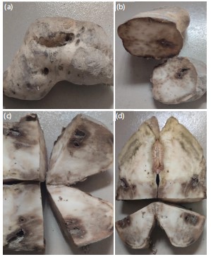
Pathogenicity test: Fresh and healthy tubers of sweet potatoes were washed with tap water and surface sterilized (70% ethanol, 2 min) and cylindrical cores (5 mm in length) were removed with a cork borer. The pure culture of each fungal isolate was introduced into the sweet potato tubers, which were then sealed with Vaseline. All treated tubers were put singly into sterile polythene bags to prevent the entry of other pathogens following the method by Kim et al.6, and incubated at room temperature for 14 days. The sweet potato tubers were cut through and examined for rot at the end of the incubation period shown in Fig. 1.
|
Macroscopic and microscopic examination: The isolated fungi were observed macroscopically by observing and studying their shape, color, size, and other morphological features.
The microscopic examination was observed by mounting the pure cultures in lacto phenol in cotton blue dye on a clean slide, well covered by a cover slide, which was observed under the microscope with a digital camera. The fungi were identified using cultural characteristics.
Statistical analysis: A Completely Randomized Design (CRD) with three replicate plates was used for the study. The data were analyzed using two-way ANOVA and the least significant differences at 5% were used to compare mean differences.
RESULTS
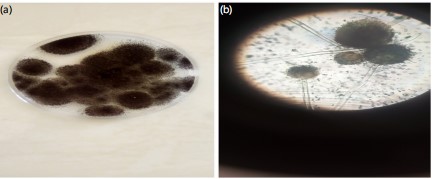
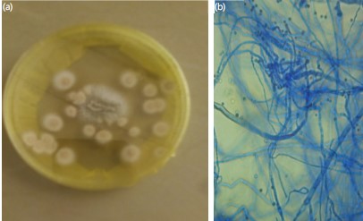
The result of this study showed that Aspergillus niger, Aspergillus flavus, Aspergillus fumigatus, Rhizopus spp., Fusarium spp., and Penicillium spp., were found associated with sweet potato rots in storage. Table 1 presents the coloration, appearance, characteristics and microscopic features of the fungi isolated from this study. Morphological characterization was used to identify the organisms. Figure 2-7 shows the different macroscopic and microscopic features of the organisms. The physical and microscopic features of the isolate were seen on the Petri dish and viewed under the microscope. The macroscopic feature of Aspergillus niger showed that the Colonies initially appeared white and later started turning black due to spore production. The texture of the colonies exhibited a woolly to granular texture due to dense conidiophore structures. The organism was rapidly growing, forming a circular, dense, and powdery colony. The surface was flat with a radial pattern. The microscopic feature of Aspergillus niger showed that hyphae is septate and branching. The conidiophores were Long and smooth. Conidia were arranged in chains, creating a characteristic “radiating head” appearance. They appeared black rough-walled, and spherical, forming radiating chains (Fig. 2).
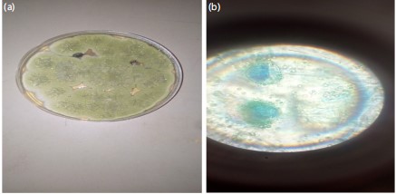
The macroscopic feature of Aspergillus flavus showed that the Colonies initially appeared grey-green, later turning dark green with age. The texture of the colonies apeared velvety to powdery due to dense conidial structures. The colony was rapidly growing, forming a circular, spreading colony with a well-defined margin. The surface was rapidly growing, forming a circular pattern. The microscopic feature of Aspergillus flavus showed that the hyphae is septate and branching. The conidiophores were Long, rough-walled, and terminate in a swollen vesicle. Conidia were yellow-green, spherical to elliptical, finely roughened, and arranged in chains. Conidia radiate around the vesicle, forming a characteristic spore head (Fig. 3).
| Table 1: | Coloration, appearance, characteristics and microscopic features of the fungi isolate | |||
| Fungi isolates | Coloration, appearance, and characteristics on PDA plates |
Microscopic features |
| Aspergillus niger | It is a black color in appearance | A microscopic view of Aspergillus niger has smooth colored conidiophores and conidia. The conidiophores are protrusions from septate and hyaline hyphae |
| Aspergillus flavus | It is a grey-green color in appearance | Conidiophores of Aspergillus flavus is colorless, thick-walled, rough, and bearing vesicles |
| Aspergillus fumigatus | It is white-yellow in appearance | Hyphae are septate and hyaline. Conidial heads are strongly columnar in an undisturbed culture. Conidiophores are smooth-walled and colourless |
| Rhizopus spp. | White-cream in color | Hyphae septate, hyaline. Conidiophores are simple or branched. Phialides grouped in brush-like clusters (penicilli) at the ends of the conidiophores. Conidia are unicellular, round or ovoid, hyaline or pigmented, rough walled or smooth in chains |
| Fusarium spp. | It is whitish-pink | Mycelium is branched, septate, hyaline or colored, inter or intracellular and uninucleate to multinucleate |
| Unknown 1 | White color with yeast-like flat appearance and formation of air mycelium |
Septate hyphae with dichotomous ramification |
| Unknown 2 | It is a white-brown color | Non septate hyphae, conidiophores are borne on the non septate sterigmata |
|
|
|
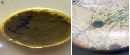
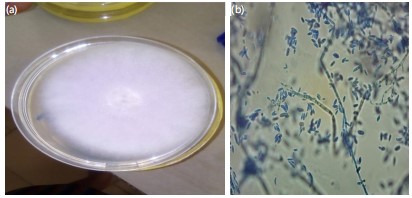
The macroscopic feature of Aspergillus fumigatus showed that the Colonies appeared whitish-yellow with a white peripheral margin. The texture of the colonies apeared velvety to powdery due to dense conidial structures. It had a dense colony with a slightly wrinkled surface, which is a rapid-growing, forming a compact. The surface is flat to slightly raised, which formed radial furrows. The microscopic feature of Aspergillus fumigatus showed that hyphae is septate, hyaline, and branching. The conidiophores were Short, smooth-walled, terminating in a small, club-shaped vesicle. Conidia were spherical, smooth to finely roughened, and arranged in long, compact chains. Conidia form columnar head (Fig. 4).
Figure 5 is the unidentified organism. The macroscopic features were a White color with yeast-like flat appearance and formation of air mycelium. The microscopic feature was Septate hyphae with dichotomous ramification.
|
|
|
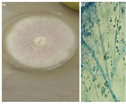
The macroscopic feature of Fusarium spp., showed that the Colonies appeared white to pale pink on the surface. The texture of the colonies appeared cottony to woolly, with aerial mycelium forming a dense, fluffy mat. It had rapidly-growing and spreading colonies with irregular margins, which was flat to slightly raised with possible radial folds. The microscopic feature of Fusarium spp., showed that hyphae is septate, transparent and branching. Conidiophores were Simple to branched, bearing conidia in clusters. Microconidia were small, oval to kidney-shaped, single-celled, and abundantly produced on short phialides. Macroconidia were sickle-shaped (fusiform), multi-celled (3-5 septa), with tapered ends, often slightly curved. Chlamydospores were thick-walled, round, resting spores, typically forming singly within the mycelium (Fig. 6).
The macroscopic feature of Rhizopus spp., showed that the Colonies appeared white at the early growth stage, later turned grayish-black due to spore production. The texture of the colonies appeared Cottony to fluffy, with a rapid spread across the growth medium. It had fast-growing, forming dense, fuzzy mycelium with black sporangia scattered across the surface, with low to moderately raised aerial hyphae giving a voluminous appearance. The microscopic features of Rhizopus spp., showed that hyphae is broad, aseptate (coenocytic), and branching. Stolons were horizontal hyphae that connected groups of sporangiophores (Fig. 7).
Table 2 presents the mean occurrence and percentage frequency of different fungal isolates responsible for various types of rot in sweet potatoes from Farin Gada Market. The key observations include: Aspergillus niger had the highest occurrence (12.00) and frequency (23.52%), making it the most prevalent fungus. Rhizopus spp., followed closely with a total occurrence of 9 and a frequency of 17.64%. Aspergillus fumigatus had a moderate occurrence (8.00) with a frequency of 15.68%. Two unidentified fungi (Unknown 1 and 2) had the lowest occurrences (2 each, 3.92%). The p-value of 0.023 indicates a statistically significant difference in the distribution of fungal isolates across different rot types.
Table 3 presents the mean occurrence and percentage frequency of different fungal isolates responsible for various types of rot in the sweet potatoes building material market. Aspergillus flavus had the highest total occurrence (9.00) and the highest frequency (19.57%). Aspergillus niger and Aspergillus fumigatus both had a total occurrence of 8.00, with frequencies of 17.39% each, showing similar prevalence in causing rot in sweet potato spoilage process. Penicillium spp., appeared moderately with a total of 7 occurrences (15.22%). Rhizopus spp., also had a total occurrence of 7.00 (15.21%). Fusarium spp., had the lowest total occurrence (4.00) with a frequency of 9.69%, making it the least frequent among the fungi in this study. Unknown 1 and 2 were identified with occurrences of 1.00 and 2.00, respectively, and frequencies of 2.71 and 4.35%, the least among the isolates. The mean occurrence rates for fungal isolates ranged from 2.71 to 19.57%. The LSD (least significant difference) values show significant differences between the isolates, with Aspergillus flavus standing out as the most frequent.
Table 4 presents the mean occurrence and percentage frequency of different fungal isolates responsible for various types of rot in sweet potatoes, Terminus Market. Aspergillus flavus had the highest total occurrence (9.00) and the highest frequency (18.00%), making it the most prevalent fungal isolate. Aspergillus niger and Aspergillus fumigatus both had a total occurrence of 8.00 (16.00% frequency each), showing their significant contribution to sweet potato spoilage. Rhizopus spp., had a total occurrence of 7.00 and a frequency of 17.64%, indicating that it is also a major contributor to spoilage. Fusarium spp., had a total occurrence of 7.00, with a frequency of 14.00%, showing a moderate level of prevalence. Penicillium spp., had a total occurrence of 4.00 (8.00% frequency), indicating lower prevalence in this market. The unidentified fungi (Unknown 1 and 2) had the lowest occurrences, with 1.00 and 2.00, corresponding to 2.00 and 4.00% frequencies, respectively. The LSD (least significant difference) value of 2.82 suggests a significant difference between fungal isolates. The p-value (<0.001) indicates a highly significant variation in the occurrence of different fungal species.
| Table 2: | Mean occurrence of each fungal isolate in Farin Gada Market | |||
| Fungal isolate | Soft rot | Black rot | Fusarium root rot | Fusarium surface rot | Total | Frequency of occurrence (%) |
| Aspergillus niger | 3.00 | 2.00 | 3.00 | 4.00 | 12.00 | 23.52±0.80a |
| Aspergillus flavus | 3.00 | 1.00 | 0.00 | 2.00 | 6.00 | 11.76±1.28c |
| Fusarium spp. | 2.00 | 0.00 | 3.00 | 2.00 | 7.00 | 13.76±1.23c |
| Penicillium spp. | 1.00 | 2.00 | 1.00 | 1.00 | 5.00 | 9.80±0.49d |
| Aspergillus fumigatus | 2.00 | 2.00 | 3.00 | 1.00 | 8.00 | 15.68±0.40bc |
| Unknown 1 | 0.00 | 0.00 | 0.00 | 2.00 | 2.00 | 3.92±0.98e |
| Unknown 2 | 0.00 | 2.00 | 0.00 | 0.00 | 2.00 | 3.92±0.98e |
| Rhizous spp. | 4.00 | 2.00 | 2.00 | 1.00 | 9 = 51 | 17.64±1.23b |
| Mean 2.68, LSD 2.84, p-value 0.023, values with same superscript are not significant | ||||||
| Table 3: | Mean occurrence of each fungal isolate in the building material market | |||
| Fungal isolate | Soft rot | Black rot | Fusarium root rot | Fusarium surface rot | Total | Frequency of occurrence (%) |
| Aspergillus niger | 3.00 | 2.00 | 2.00 | 1.00 | 8.00 | 17.39±0.88ab |
| Aspergillus flavus | 3.00 | 2.00 | 2.00 | 2.00 | 9.00 | 19.57±0.54a |
| Penicillium spp. | 2.00 | 0.00 | 3.00 | 2.00 | 7.00 | 15.22±1.37b |
| Fusarium spp. | 2.00 | 1.00 | 0.00 | 1.00 | 4.00 | 9.69±0.92c |
| Aspergillus fumigatus | 3.00 | 2.00 | 2.00 | 1.00 | 8.00 | 17.39±0.89ab |
| Unknown 1 | 0.00 | 0.00 | 0.00 | 1.00 | 1.00 | 2.71±0.68d |
| Unknown 2 | 0.00 | 2.00 | 0.00 | 0.00 | 2.00 | 4.35±1.09d |
| Rhizopus spp. | 4.00 | 2.00 | 1.00 | 0.00 | 7.00 = 46 | 15.21±1.86b |
| Mean 3.89, LSD 3.12, p<0.001, values with same superscript are not significant | ||||||
| Table 4: | Mean occurrence of each fungal isolate in Terminus Market | |||
| Fungal isolate | Soft rot | Black rot | Fusarium root rot | Fusarium surface rot | Total | Frequency of occurrence (%) |
| Aspergillus niger | 3.00 | 2.00 | 2.00 | 1.00 | 8.00 | 16.00±0.82ab |
| Aspergillus flavus | 3.00 | 2.00 | 2.00 | 2.00 | 9.00 | 18.00±0.50a |
| Fusarium spp. | 2.00 | 0.00 | 3.00 | 2.00 | 7.00 | 14.00±1.26b |
| Penicillium spp. | 2.00 | 1.00 | 0.00 | 1.00 | 4.00 | 8.00±0.82c |
| Aspergillus fumigatus | 3.00 | 2.00 | 2.00 | 1.00 | 8.00 | 16.00±0.82ab |
| Unknown 1 | 0.00 | 0.00 | 0.00 | 1.00 | 1.00 | 2.00±0.50d |
| Unknown 2 | 0.00 | 2.00 | 0.00 | 0.00 | 2.00 | 4.00±1.00d |
| Rhizopus spp. | 4.00 | 2.00 | 1.00 | 0.00 | 7.00 = 50 | 17.64±1.23b |
| Mean 2.66, LSD 2.82, p<0.001, values with same superscript are not significant | ||||||
| Table 5: | Percentage frequency of occurrence of fungi isolated from 3 markets | |||
| Organisms | Farin Gada | Building material | Terminus | Total | Frequency of occurrence (%) |
| Aspergillus niger | 12.00 | 13.00 | 8.00 | 33.00 | 22.44±1.79a |
| Aspergillus flavus | 6.00 | 9.00 | 9.00 | 24.00 | 16.32±1.18b |
| Fusarium spp. | 7.00 | 11.00 | 7.00 | 25.00 | 17.00±1.57b |
| Penicillium spp. | 5.00 | 4.00 | 4.00 | 13.00 | 8.84±0.39d |
| Aspergillus fumigatus | 8.00 | 8.00 | 8.00 | 24.00 | 16.33±0.00b |
| Unknown 1 | 2.00 | 0.00 | 1.00 | 3.00 | 2.04±0.68e |
| Unknown 2 | 2.00 | 2.00 | 2.00 | 6.00 | 4.08±0.00e |
| Rhizopus spp. | 9.00 | 3.00 | 7.00 | 19.00 | 12.93±2.08c |
| Mean | 5.67 | ||||
| LSD | 2.00 | ||||
| p-value | <0.0001 | ||||
| At p≤0.05 there was a significant difference in the frequency of occurrence of the organism. Values are presented as Mean±Standard Error of Mean. Ranking is done across fungal isolates and values with same superscript are not significant | |||||
Table 5 shows that Aspergillus niger had the highest total occurrence (33.00) and the highest frequency (22.44%), making it the most prevalent fungal species across the three markets. Penicillium spp., had a total occurrence of 13.00 (8.84% frequency), indicating a relatively lower contribution to spoilage. The unidentified fungi (Unknown 1 and 2) had the lowest occurrences (3.00 and 6.00, respectively), with frequency values of 2.04 and 4.08%, suggesting minimal impact on sweet potato spoilage.
| Table 6: | Pathogenicity test of the organisms isolated from sweet potatoes | |||
| Organisms | Weight of tuber | Weight of 7 days | Weight lost (%) | Diameter of rot |
| Aspergillus niger | 116.70±11.0 | 70.58±8.05 | 34.63±3.85 | 16.80±1.20 |
| Aspergillus flavus | 118.37±31.62 | 68.87±26.74 | 43.86±7.59 | 28.30±0.70 |
| Rhizopus spp. | 116.73±11.13 | 94.26±11.53 | 19.19±2.48 | 12.45±0.65 |
| Aspergillus fumigatus | 148.42±31.26 | 118.34±24.86 | 20.77±0.46 | 15.45±1.15 |
| Penicillium spp. | 110.12±21.45 | 85.94±14.19 | 21.53±2.45 | 11.35±0.35 |
| Fusarium spp. | 110.12±21.45 | 61.00±2.70 | 42.65±4.84 | 1s9.95±0.75 |
| Unknown 1 | 154.4±323.01 | 125.344±7.89 | 17.77±7.14 | 9.00±1.20 |
| Unknown 2 | 96.66±11.94 | 75.76±24.03 | 21.87±1.76 | 14.95±0.80 |
| LSD | 19.41 | |||
| p-value | <0.001 | |||
| at p≤0.05, there was a significant difference in the percentage weight lost and the diameter of rot. Values were presented as Mean±Standard Deviation, values with same superscript are not significant | ||||
Table 6 shows the pathogenicity test of fungal isolates on sweet potatoes. Weight loss (%) as an Indicator of fungal pathogenicity: Aspergillus flavus caused the highest weight loss (43.86%), making it the most aggressive pathogen in terms of tuber degradation. Fusarium spp., also induced significant weight loss (42.65%), indicating high pathogenicity. Aspergillus niger caused 34.63% weight loss, showing moderate pathogenicity. Rhizopus spp., had the lowest weight loss (19.19%), suggesting it is less aggressive compared to Aspergillus and Fusarium species. Diameter of Rot (cm): Another measure of pathogenicity. Aspergillus flavus had the largest rot diameter (28.30 cm), further confirming its strong ability to cause extensive decay. Fusarium spp., followed with a rot diameter of 19.95 cm. Aspergillus niger and Aspergillus fumigatus produced moderate rot diameters of 16.80 and 15.45 cm, respectively. Unknown 1 caused the smallest rot diameter (9.00 cm), indicating the lowest pathogenic impact. The p-value (<0.001) indicates that the differences observed are statistically significant, reinforcing the variability in fungal pathogenicity.
DISCUSSION
The result of this study showed that Aspergillus niger, Aspergillus flavus, Aspergillus fumigatus, Rhizopus spp., Fusarium spp., Penicillium spp., were found associated with sweet potato rots in storage. The isolated fungi and their percentage of occurrence include Aspergillus niger (22.45%) as the highest frequency followed by Fusarium spp. (17.01%) followed by Aspergillus flavus and Aspergillus fumigatus both have (16.33%) followed by Rhizopus spp., and (12.93%), Penicillium spp. (8.84%) Several researchers have also documented the microbes that are responsible for sweet potato rotting during the post-harvest period.
In this research, Aspergillus niger was found to be the most predominant fungi isolates with 21.67% it has the highest percentage occurrence and this is similar to the report of Tortoe et al.14, that Aspergillus niger and Aspergillus flavus to be abundant fungal species during post-harvest storage of sweet potato.
The results obtained from this research agree with the findings of Sila et al.15, who identified Aspergillus spp., Fusarium spp., and Rhizopus spp., as some of the fungi that causes spoilage of sweet potato. Aspergillus fumigatus, known as the sweet potato pathogen16,17. Pathogenicity test revealed that four of the isolated fungi were highly pathogenic, Aspergillus flavus, and Fusarium spp., induced the most extensive rots, Aspergillus niger, Aspergillus fumigatus and were moderately pathogenic while Rhizopus spp. Pathogenicity test revealed that four of the isolated fungi were highly pathogenic, Aspergillus flavus, and Fusarium spp., induced the most extensive rots, Aspergillus niger, Aspergillus fumigatus and were moderately pathogenic while Rhizopus spp., Penicillium spp., are least pathogenic.
This study confirmed that fungi are the primary cause of sweet potato post-harvest diseases during storage, significantly reducing its market availability and nutrition. The findings indicate that Aspergillus species were the most prevalent cause of the sweet potato tubers diseases in storage. These fungal diseases contribute to large post-harvest losses notwithstanding the crop’s economic and nutritional importance. The presence of Aspergillus niger was particularly alarming causing black rot disease. Additionally, most of the identified fungi demonstrated the ability to rapidly reinfect healthy tubers, creating economic challenges for sweet potato sellers in Jos, Plateau State, Nigeria. This agrees with the study of Oduola et al.18.
The rapid deterioration of sweet potatoes is often aggravated by exposure to environmental conditions and mechanical damage sustained during handling and transportation. To reduce post-harvest diseases, several preventive measures should be implemented, including careful handling of tubers, proper cleaning of storage and transit containers, minimizing physical injuries, and maintaining hygienic practices among handlers. Providing suitable storage facilities is also crucial. Furthermore, adopting cost-effective disease management strategies that are accessible to small-scale farmers is essential to ensuring that sweet potato remains a viable contributor to food security and the national economy.
According to this study, the main cause of sweet potato disease during storage is fungi, specifically Aspergillus niger and Aspergillus flavus to be abundant fungal species during post-harvest storage of sweet potato. This agrees with the findings of Paul et al.16.
Pathogenicity tests verified that this fungus may infect healthy tubers again, resulting in large financial losses and possible health hazards from the formation of mycotoxin.
CONCLUSION
The findings highlight the major fungal pathogens responsible for sweet potato spoilage in storage at the different markets, with Aspergillus flavus and Fusarium spp., posing the highest threat. The results highlight the necessity of better postharvest handling, clean storage conditions, and reasonably priced disease management strategies for farmers. Future studies should focus on developing biocontrol strategies to mitigate post-harvest losses and improve sweet potato storage conditions. To improve food security and lower postharvest losses, future research should concentrate on creating bio-based antifungal therapies and investigating resistant sweet potato cultivars. Governments should provide proper storage facilities for sweet potatoes and other essential tubers to ensure national food security. Additionally, careful handling during harvest is crucial to prevent bruises and mechanical damage, which significantly contribute to post-harvest losses.
SIGNIFICANCE STATEMENT
By identifying key pathogens and their pathogenicity, this research highlights the need for improved storage practices and effective disease management strategies. The findings contribute to the development of sustainable and affordable postharvest handling techniques, supporting smallholder farmers, traders, and policymakers in reducing losses and ensuring a more stable food supply. This study offers important insights into the fungal pathogens that cause sweet potato spoilage during storage, with Aspergillus spp., being the most common. They also highlight the economic and food security implications of postharvest losses, highlighting the risks posed to consumer health by mycotoxin-producing fungi.
REFERENCES
- Laurie, S.M., J. Mulabisana, R. Sutherland and D. Sivakumar et al., 2024. Seventy years of sweet potato [Ipomoea batatas L. (LAM)] research in South Africa. Crop Sci., 64: 1112-1128.
- Adeyonu, A.G., O.L. Balogun, B.O. Ajiboye, I.B. Oluwatayo and A.O. Otunaiya, 2019. Sweet potato production efficiency in Nigeria: Application of data envelopment analysis. AIMS Agric. Food, 4: 672-684.
- Rees, D., R. Kapinga and van Quirien Oirschot, 2003. Sweet Potato Post-Harvest Assessment: Experiences from East Africa. Natural Resources Institute, UK, ISBN: 9780859545488, Pages: 129.
- Agu, K.C., G.U. Nweke, N.S. Awah, B.C. Okeke, I.C.C. Mgbemena, R.N. Okigbo and U.C. Ngenegbo, 2015. Fungi associated with the post-harvest loss of sweet potato. Int. J. Res. Stud. Biosci., 3: 33-38.
- Beckley, F. and S.O. Awoyemi, 2021. Occurrence of postharvest fungal rots of sweet potato (Ipomoea batatas (L) Lam.) in Southwest Nigeria and their control with sawdust extracts. J. Exp. Agric. Int., 43: 24-33.
- Kim, S., T.H. Kim, M.N. Chung, Y.H. Lee, I.B. Lee, H.U. Lee and W. Park, 2022. Incidence rates of root rot in sweetpotato caused by cultivation soil and soil microorganisms during storage periods. Front. Plant Sci., 13.
- Parmar, A., O. Hensel and B. Sturm, 2017. Post-harvest handling practices and associated food losses and limitations in the sweetpotato value chain of Southern Ethiopia. NJAS: Wageningen J. Life Sci., 80: 65-74.
- Udoh, I.P., C.I. Eleazar, B.O. Ogeneh and M.E. Ohanu, 2015. Studies on fungi responsible for the spoilage/deterioration of some edible fruits and vegetables. Adv. Microbiol., 5: 285-290.
- Nath, B., G. Chen, C.M. O’Sullivan and D. Zare, 2024. Research and technologies to reduce grain postharvest losses: A review. Foods, 13.
- Olaitan, O.O., 2012. Bio-deterioration of sweet potato (Ipomoea batatas Lam) in storage, inoculation-induced quality changes, and control by modified atmosphere. J. Applied Sci. Environ. Manage., 16: 189-193.
- Alegbeleye, O., O.A. Odeyemi, M. Strateva and D. Stratev, 2022. Microbial spoilage of vegetables, fruits and cereals. Appl. Food Res., 2.
- Prusky, D.B. and E. Sionov, 2021. Special Issue "Interplay between fungal pathogens and harvested crops and fruits". Microorganisms, 9.
- Barnett, H.L. and B.B. Hunter, 1998. Illustrated Genera of Imperfect Fungi. 4th Edn., APS Press, St. Paul, Minnesota, ISBN: 978-0-89054-192-0, Pages: 218.
- Tortoe, C., M. Obodai and W. Amoa-Awua, 2010. Microbial deterioration of white variety sweet potato (Ipomoea batatas) under different storage structures. Int. J. Plant Biol., 1.
- Sila, M.D., C.I.C. Ogbonna, D.D. Nyam, P.A. Wuyep and V.M. Shutt, 2021. Studies on the fungal isolates of the soil spots of sweet potato (Ipomoae batatas (L.) Lam) root tubers in the field. Aust. J. Basic Appl. Sci., 15: 1-9.
- Paul, N.C., S. Park, H. Liu, J.G. Lee, G.H. Han, H. Kim and H. Sang, 2021. Fungi associated with postharvest diseases of sweet potato storage roots and in vitro antagonistic assay of Trichoderma harzianum against the diseases. J. Fungi, 7.
- Gyasi, E., S. Akrofi, B.A. Adongo, E.A. Osafo, D.A. Kotey and A. Mohammed, 2022. Fungi associated with sweet potato tuber rot at CSIR-PGRRI, Bunso, Eastern Region, Ghana. Ghana J. Agric. Sci., 57: 106-112.
- Oduola, A.A., K.O. Awojobi and S.M. Adeyemo, 2018. Microorganisms associated with post harvest spoilage of sweet potatoes in Ile-Ife, Nigeria. J. Microbiol. Res., 8: 1-8.
How to Cite this paper?
APA-7 Style
Adaku,
N.I., Yoro,
M.S., Nwadiaro,
P.O. (2025). Post-Harvest Fungal Diseases of Stored Sweet Potatoes (Ipomoea batatas (L.) Lam) in Three Markets of Jos, Plateau State. Asian Journal of Plant Pathology, 19(1), 16-26. https://doi.org/10.3923/ajpp.2025.16.26
ACS Style
Adaku,
N.I.; Yoro,
M.S.; Nwadiaro,
P.O. Post-Harvest Fungal Diseases of Stored Sweet Potatoes (Ipomoea batatas (L.) Lam) in Three Markets of Jos, Plateau State. Asian J. Plant Pathol. 2025, 19, 16-26. https://doi.org/10.3923/ajpp.2025.16.26
AMA Style
Adaku
NI, Yoro
MS, Nwadiaro
PO. Post-Harvest Fungal Diseases of Stored Sweet Potatoes (Ipomoea batatas (L.) Lam) in Three Markets of Jos, Plateau State. Asian Journal of Plant Pathology. 2025; 19(1): 16-26. https://doi.org/10.3923/ajpp.2025.16.26
Chicago/Turabian Style
Adaku, Nnebechukwu,, Ijeoma, Musa Stephanie Yoro, and P. O. Nwadiaro.
2025. "Post-Harvest Fungal Diseases of Stored Sweet Potatoes (Ipomoea batatas (L.) Lam) in Three Markets of Jos, Plateau State" Asian Journal of Plant Pathology 19, no. 1: 16-26. https://doi.org/10.3923/ajpp.2025.16.26

This work is licensed under a Creative Commons Attribution 4.0 International License.