
New Records and Host Plant Associations of Erysiphe Species in Pakistan Based on Morpho-Anatomical and Molecular Characterization
-
Muhammada Jabeen
Institute of Botany, University of the Punjab, Lahore, Pakistan
Najam ul Sehar AfshanInstitute of Botany, University of the Punjab, Lahore, Pakistan
Muskan AmjadInstitute of Botany, University of the Punjab, Lahore, Pakistan
Arooma Saleem
Institute of Botany, University of the Punjab, Lahore, Pakistan
Ayesha ZahidInstitute of Botany, University of the Punjab, Lahore, Pakistan
Saliha AfzalInstitute of Botany, University of the Punjab, Lahore, Pakistan
Abdul Nasir KhalidInstitute of Botany, University of the Punjab, Lahore, Pakistan
| Received 23 May, 2025 |
Accepted 10 Sep, 2026 |
Published 20 Sep, 2026 |
Background and Objective: Powdery mildew caused by Erysiphe species is widely distributed and affects a broad range of host plants, yet comprehensive records from Pakistan remain limited. The objective of this study was to investigate powdery mildew-infected plants collected from different Regions of Pakistan and to identify the associated Erysiphe species using an integrated morpho-anatomical and molecular approach, to document new host records and distribution reports within the country. Materials and Methods: Mycological field surveys were conducted across various regions of Pakistan from 2020 to 2024. Plant specimens exhibiting powdery mildew symptoms were collected and examined through detailed morpho-anatomical characterization. Molecular phylogenetic analyses were performed to confirm species identity using an integrated taxonomic approach. Results: The infected plants showed typical powdery mildew symptoms, including white powdery growth on foliage and small black to brownish rounded ascomata. Aquilegia fragrans, Lantana indica and Ranunculus repens were confirmed as new host records for Erysiphe aquilegiae. Furthermore, Erysiphe lespedezae is reported for the first time on Lespedeza tomentosa in Pakistan. Conclusion: The study expands the known host range and distribution of Erysiphe species in Pakistan and highlights the importance of combining morpho-anatomical and molecular approaches for accurate fungal identification.
INTRODUCTION
Powdery mildew fungi are obligate biotrophic parasites that establish long-term associations with living host cells1. These pathogens infect various parts of angiosperms including leaves, stems, flowers and fruits by forming characteristic white, powdery films on host surfaces. Globally, 16 genera and 904 species of powdery mildews have been documented2. In Pakistan, 44 species have been reported to date, with the genus Erysiphe being the most prevalent. This genus infects 55 host species across 20 plant families, including members of the Salicaceae family3. Similarly, members of the Fabaceae and Ranunculaceae family are also susceptible to powdery mildew infections. During fungal surveys conducted between 2020 and 2024 in various Regions of Pakistan including Fairy Meadows (Gilgit Baltistan), Nathiagali, Shogran, Mansehra (Khyber Pakhtunkhwa) and Jhelum valley (Azad Jammu and Kashmir) and Leepa valley. Powdery mildew infections were consistently observed on multiple host plants, including Aquilegia fragrans, Lantana indica, Lespedeza tomentosa and Ranunculus repens. Morpho-anatomical and phylogenetic analyses identified the causative fungi as different species of the widespread and diverse genus Erysiphe. These newly collected Erysiphe species from different localities contribute to our understanding of plant-fungus interactions and indicate an expansion in host range. The primary objectives of this study are to identify and confirm the identity of the fungal species infecting newly recorded host plants using both morphological and molecular approaches, to compare these findings with previously reported species and to discuss possible environmental, ecological and evolutionary factors contributing to host expansion. Previously, Erysiphe aquilegiae has been reported on Aquilegia vulgaris, A. fragrans, Delphinium ajacis and Ranunculus repens4 while from Pakistan, it is reported on Clematis montana5. However, in the present study, these host species are newly recorded for E. aquilegiae in Pakistan. Similarly, Erysiphe lespedezae was previously reported only on Kummerowia striata in Korea. This study marks the first report of this species from Pakistan.
MATERIALS AND METHODS
Description of sampling sites: Specimens were collected from Fairy Meadows, Jhelum, Nathia Gali, Shogran, Leepa Valley and Mansehra, during 2020 to 2024.
Collection and preservation of specimens: Collected plant samples were shade-dried using blotting paper. Diseased specimens were pressed and preserved following standard herbarium techniques. Leaves exhibiting powdery mildew symptoms were stored in paper envelopes containing detailed collection information and sealed in airtight plastic bags to prevent contamination. All voucher specimens were deposited in the herbarium of the Institute of Botany, University of the Punjab, Lahore (LAH). Infected plant samples were photographed in the field using an EMZ-TR stereomicroscope (Meiji Techno, Japan). For microscopic examination, fungal structures were mounted in lactic acid and analyzed under a LABOMED light microscope. Morphological features including chasmothecia, appendages, asci, ascospores, conidiophores, conidia and appressoria were measured using Scope Image 9.0 (X5) camera software.
DNA extraction and PCR amplification: Genomic DNA was extracted from infected plant tissues using the GeneJET Plant Genomic DNA Purification Mini Kit (Thermo Fisher Scientific, Waltham, MA, USA) following the manufacturer’s protocol. For molecular identification, PCR amplification of the internal transcribed spacer (ITS) region of nuclear ribosomal DNA (ITS-nrDNA) was performed using the primer pair PMITS1 (forward: 5'-TCGGACTGGCCyAGGGAGA-3') and PMITS2 (reverse: 5'-TCACTCGCCGTTACTGAGGT-3') as described by Cunnington et al.6. PCR products were separated by electrophoresis on a 1% agarose gel stained with ethidium bromide and visualized under UV light7. Sanger sequencing of purified amplicons was carried out by BGI (Hong Kong, China) and TsingKe Biological Technology (China). The newly generated ITS sequences were submitted to the NCBI GenBank database.
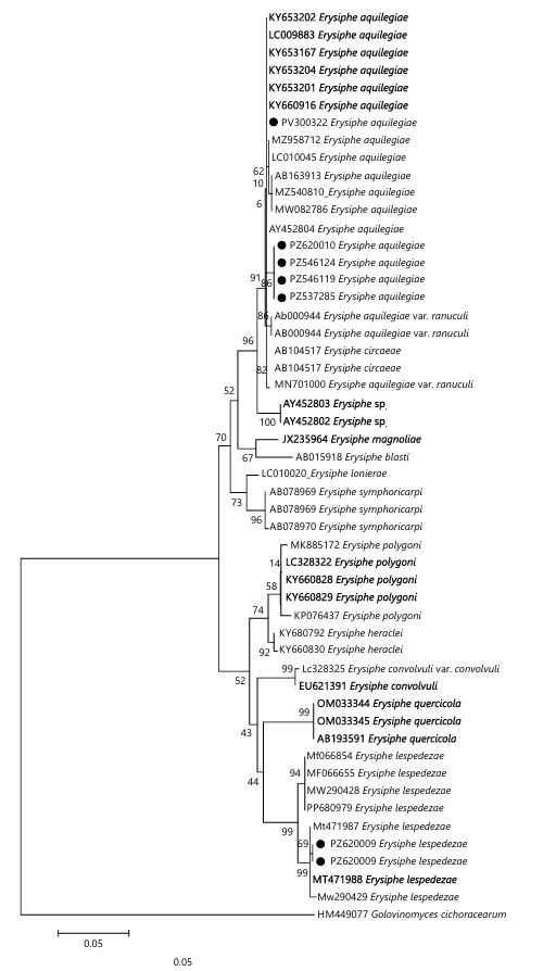
Sequence alignment and phylogenetic analysis: The internal transcribed spacer (ITS) region of each specimen was amplified and sequenced. Forward and reverse sequences were assembled and edited using BioEdit Sequence Alignment Editor8. Nucleotide similarity searches were conducted using BLAST at the National Center for Biotechnology Information (NCBI) to identify closely related taxa. The most similar sequences retrieved from GenBank were selected for phylogenetic analysis. Multiple sequence alignment was performed with MAFFT v7.0 using default settings and the alignment was manually trimmed at the 5' and 3' ends to remove poorly aligned or ambiguous regions, retaining only conserved sites. The final dataset comprised 583 nucleotide positions, including 427 conserved sites, 150 variable sites, 87 parsimony-informative sites and 63 singleton variants. Phylogenetic relationships were inferred using the Maximum Likelihood (ML) method in MEGA 6.09. The Kimura 2 parameter was selected based on model testing for best fit to the nucleotide substitution pattern. Golovinomyces cichoracearum (HM449077) was used as the outgroup to root the tree. The resulting phylogenetic tree demonstrated that the specimens clustered with Erysiphe aquilegiae, consistent with morphological identification.
|
RESULTS AND DISCUSSION
Taxonomy
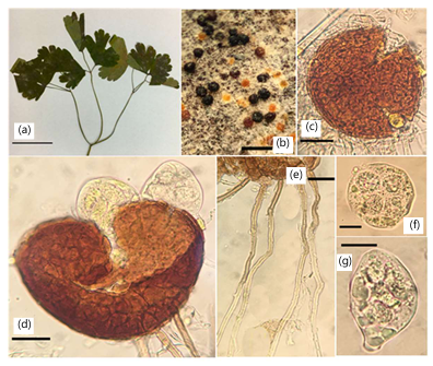
Erysiphe aquilegiae DC., Fl. franç., Edn 3 (Paris) 5/6: 105 (1815). Fig 1(a-g)
Specimens examined: On Aquilegia fragrans Benth. (Ranunculaceae), Pakistan, Khyber Pakhtunkhwa, Nathiagali, 2,410 m.a.s.l., 20 August, 2020, S. Afzal & N. S. Afshan (PM-34). (LAH04026; GenBank no. PZ546119) Fig. 1(a-g).
On Aquilegia fragrans Benth. (Ranunculaceae), Pakistan, Khyber Pakhtunkhwa, Mansehra, Shogran, 2,362 m.a.s.l., 29 September, 2021, S. Afzal & N. S. Afshan (PM-39).( LAH04326; GenBank no. PZ537285).
On the leaves of Ranunculus repens Stephan. Ex Willd. (Ranunculaceae), with both of the anamorphic stage and telomorphic stages, Pakistan, Gilgit Baltistan, District Diamer, Fairy Meadows, at 3,300 m.a.s.l., August 11, 2023, collected by Najam ul Sehar Afshan and Muhammada jabeen (FM-10). (LAH04426; GenBank no. PZ546124).
Description
Mycelium: Amphigenous, thick, persistent.
|
| Hyphae | : | Septate, hyaline, thick-walled, smooth to almost so, 4-7 μm wide. | |
| Chasmothecia | : | Scattered or often gregarious, globose, light to dark chocolate brown, (99-)101-111(-117) μm in diameter, Fig. 1d. | |
| Peridium | : | With thick, fairly conspicuous layers, irregularly shaped, compactly arranged, light to dark brown, 10-19 μm in diameter. | |
| Appendages | : | Usually numerous, about 10-40 in the lower half, mycelioid, simple, brown throughout or paler toward the apex, septate, wall thin, smooth to rough (127-)137-210(-266)×(06-)07-08(-10) μm, Fig. 1e. | |
| Asci | : | Bitunicate, broadly ellipsoid-obovoid, saccate, (59-)60-66(-69)×(39-)43-45(-47) μm, sessile, rarely short stalked, 4 spored, Fig. 1f-g. |
|
Ascospores: Ellipsoid-ovoid, colorless, (14-)15-17(-18)×(8-)9-10(-11) μm.
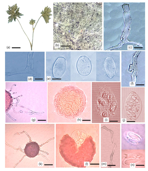
First record on Ranunculus repens (Ranunculaceae) in Pakistan Fig. 2a-n
Specimens examined: On the leaves of Ranunculus repens Stephan. ex Willd. (Ranunculaceae), with teleomorphic stage, Pakistan, Azad Jammu and Kashmir, Jhelum Valley, at 3, 200 m.a.s.l., August 5, 2024, collected by Najam ul Sehar Afshan (LP-21) (LAH03926: GenBank no. PZ620009).
Additional material examined: On the leaves of Lantana indica Wall. (Verbenaceae), with only the anamorphic stage, Pakistan, Gilgit Baltistan, District Diamer, Fairy Meadows, at 3,300 m.a.s.l., August 11, 2023, N. S., Afshan and M. Jabeen (BAT-01) (LAH38679, Genbank no. PV300322).
Description
| Mycelium | : | Amphigenous, dense, persistent, pannose. | |
| Hyphae | : | Hyaline, thick-walled, smooth, septate. | |
| Appressoria | : | Slightly nipple-shaped, 7 μm, Fig. 2c. | |
| Conidiophores | : | Hyaline, straight, short, 54-71×7-19 μm, Fig. 2f. | |
| Foot cell | : | Straight, followed by 1-2 cells, 14-19×7-11 μm, Fig. 2d. |
|
| Conidia | : | Cylindrical, thin-walled, 29-32×11-20 μm, Fig. 2e. | |
| Germinating conidia | : | Euoidium type. | |
| Chasmothecia | : | Scattered, globose, black to red when mature, abundant, 112-185 μm in diameter, Fig. 2g. | |
Peridial cells |
: | Thick, compact, polygonal, inconspicuous, 6-16×5-11 μm. | |
Appendages |
: | Long, numerous, brown, septate, thin, smooth-walled, covering the entire surface of the fruiting body, 47-154×5-8 μm, Fig. 2m. | |
Asci |
: | Numerous, stalked, thick-walled, containing 5-6 ascospores, measuring up to 64 μm in length and 37 μm in width, Fig. 2l. | |
| Ascospores | : | Ellipsoid, green in color, 14-26×8-11 μm, Fig. 2n. |
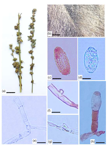
Erysiphe lespedezae R.Y. Zheng & U. Braun, Mycotaxon 18(1): 142, 1983 Fig. 3a-h
Specimens examined: Erysiphe lespedezae on the leaves of Lespedeza tomentosa Thunb. (Fabaceae), with anamorphic stage, Pakistan, Azad Jammu and Kashmir, Leepa valley, 3000 m.a.s.l, August 21, 2024, N.S. Afshan (LP-38) (LAH04226) GenBank no. PZ546106.
On the leaves of Lespedeza tomentosa Thunb. (Fabaceae), with anamorphic stage, Pakistan, Gilgit Baltistan, Fairy Meadows, 3000 m.a.s.l, August 21, 2024, N.S. Afshan (FM-26 LAH04226: GenBank no. PZ620010).
Description
| Mycelium | : |
Amphigenous, occurring in irregular patches. | |
| Hyphae | : |
Hyaline, thin-walled and septate. | |
| Conidiophores | : |
Long, erect, arising centrally from the top of the mother cell, 47-83×3-6 μm, Fig. 3h. | |
| Foot cells | : |
Cylindrical, about 15-35×5-9 μm, Fig. 3e. Followed by 2-3 cells that are almost equal in length, forming conidia singly. | |
| Conidia | : | Ellipsoid-ovoid, 25-45×15-25 μm, Fig. 3c-d. |
Appressoria slightly swollen, with lobed structures, Fig. 3f-g.
Molecular phylogeny: The ITS region of seven specimens as mentioned in Fig. 4, with accession numbers (PZ537285, PZ546119, PZ546124, PZ546106, PV300322, PZ620009, PZ620010) was amplified and sequenced. Sequence similarity searches were performed using BLAST against GenBank. Phylogenetic analysis was conducted using the Maximum Likelihood method based on the Kimura 2-parameter model with 100 bootstrap replicates. Golovinomyces cichoracearum (HM449077) was used as an outgroup. The aligned dataset comprised 583 nucleotide positions, including 427 conserved, 150 variable and 87 parsimony-informative sites. The five newly generated Erysiphe aquilegiae isolates (PV300322, PZ620010, PZ546124, PZ546119 and PZ537285) clustered together in a well-supported monophyletic clade with 91% bootstrap support. This clade was recovered as the sister lineage to E. aquilegiae var. ranunculi (AB000944 and MN701000) with 86% bootstrap support and was closely related to E. circaeae (AB104517) with 96% bootstrap support, confirming the phylogenetic placement of the newly generated isolates within the E. aquilegiae species complex. The two newly generated Erysiphe lespedezae isolates (PZ620009 and PZ546106) clustered together in a distinct subclade with 69% bootstrap support. This subclade was sister to the reference isolate MT471987 with 99% bootstrap support and grouped with other E. lespedezae reference isolates (MT471986, MF066654, MW290428, PP680979, MW290429 and MF066655), confirming the phylogenetic placement of the newly generated isolates within E. lespedezae.
DISCUSSION
The identification of Erysiphe aquilegiae in the present study is supported by both morphological characteristics and molecular phylogenetic analysis. The observed morphological features, particularly the structure of chasmothecia, appendages, asci and ascospores are consistent with previously published descriptions of the species. Molecular data further support this identification, as the isolates clustered with authenticated sequences of E. aquilegiae with strong bootstrap support.
Erysiphe aquilegiae is a widely distributed species reported on numerous hosts, primarily within the Ranunculaceae, across North America, Europe, Asia, Australia, New Zealand, South Africa and South America. Previous studies have demonstrated that morphologically similar collections from unrelated hosts may also be assigned to this species based on diagnostic features. For example, Bolay (2005) assigned anamorphic collections on Swertia perennis (Gentianaceae) and both anamorphic and teleomorphic collections on Magnolia liliiflora (Magnoliaceae) to E. aquilegiae due to their conforming morphology1. In Pakistan, E. aquilegiae has previously been reported on Thalictrum elegans10 and Clematis montana11. The present study extends its host range by documenting its occurrence on Aquilegia fragrans and Ranunculus repens, which represent new host records for the country. The occurrence of this species on diverse hosts and across multiple ecological regions indicates a broader host adaptability and distribution.
Similarly, Erysiphe lespedezae was identified based on characteristic morphological features, including conidiophore structure, foot-cell morphology and conidial shape, which are consistent with the original description of the species. Molecular identification using ITS sequence data further confirmed its placement within E. lespedezae, clustering with previously reported sequences from Taiwan (MT471986, MT471987, MW290429). This species was first reported from Korea12 and has subsequently been recorded on Bauhinia variegata, B. blakeana and Desmodium caudatum in Taiwan13. The present study documents E. lespedezae on Lespedeza tomentosa, which is consistent with its known host range within the Fabaceae. Importantly, this represents the first report of Erysiphe lespedezae from Pakistan, thereby extending its known geographical distribution. The combined use of morphological and molecular data provides strong evidence for accurate species identification and highlights new host associations and distributional records of powdery mildew fungi in Pakistan.
CONCLUSION
In this study, new records of Erysiphe species on various host plants from different regions of Pakistan are reported and described based on morphological, anatomical and phylogenetic analyses. Among these, a new record of an Erysiphe species is documented for the first time in Pakistan. This research highlights the diversity, distribution and host range of Erysiphe species, contributing to the documentation of fungal biodiversity and supporting improved disease management strategies in the country’s natural vegetation. Morphological observations combined with ITS rDNA sequencing have confirmed these new host and species records.
SIGNIFICANCE STATEMENT
This study provides important new information on the diversity, host associations and distribution of Erysiphe species in Pakistan. By integrating morpho-anatomical observations with molecular characterization, the study strengthens the reliability of species identification and confirms previously undocumented host associations. The identification of new host records for Erysiphe aquilegiae and the first record of Erysiphe lespedezae on Lespedeza tomentosa in Pakistan expand the current knowledge of powdery mildew-host relationships and contribute valuable baseline information for fungal biodiversity, taxonomy and future plant disease surveillance in the region.
ACKNOWLEDGMENT
We pay sincere thanks to Prof. Dr. Abdul Rehman Niazi, Institute of Botany, University of the Punjab, Lahore for his help during this research work.
REFERENCES
- Hückelhoven, R. and R. Panstruga, 2011. Cell biology of the plant-powdery mildew interaction. Curr. Opin. Plant Biol., 14: 738-746.
- Riaz, M., N.U.S. Afshan, S. Afzal, I. Zafar, A. Saleem and A.N. Khalid, 2024. DNA barcoding and scanning electron microscopy reveals Erysiphe ahmadii sp. nov. and a new record Erysiphe populicola (Erysiphaceae, Helotiales) on salicaceous hosts from Pakistan. Microsc. Res. Tech., 87: 21-30.
- Yeh, Y.W., M. Piepenbring and R. Kirschner, 2025. Twenty-nine new host records of powdery mildew fungi (Erysiphaceae) in Taiwan with an updated checklist. Bot. Stud., 66.
- Braun, U. and R.T.A. Cook, 2012. Taxonomic Manual of the Erysiphales (Powdery Mildews). CBS-KNAW Fungal Biodiversity Centre, Netherlands, ISBN: 9789070351892, Pages: 707.
- Afshan, N.S. and M. Riaz, 2022. First record of Erysiphe aquilegiae on Clematis Montana (Himalayan Clematis) from Pakistan. Nova Hedwigia, 114: 69-76.
- Cunnington, J.H., S. Takamatsu, A.C. Lawrie and I.G. Pascoe, 2003. Molecular identification of anamorphic powdery mildews (Erysiphales). Australas. Plant Pathol., 32: 421-428.
- Stöger, A., J. Schaffer and W. Ruppitsch, 2006. A rapid and sensitive method for direct detection of Erwinia amylovora in symptomatic and asymptomatic plant tissues by polymerase chain reaction. J. Phytopathol., 154: 469-473.
- Hall, T.A., 1999. BioEdit: A user-friendly biological sequence alignment editor and analysis program for windows 95/98/NT. Nucleic Acid Symp. Ser., 41: 95-98.
- Tamura, K., G. Stecher and S. Kumar, 2021. MEGA11: Molecular evolutionary genetics analysis version 11. Mol. Biol. E, 38: 3022-3027.
- Aman, N., A.N. Khalid and J.M. Moncalvo, 2022. A compendium of macrofungi of Pakistan by ecoregions. MycoKeys, 89: 171-233.
- Afshan, N.U.S., M. Riaz, N. Liaqat, J. Majeed and M. Jabeen et al., 2022. Erysiphe pakistanica (Erysiphaceae), a new species on Indian willows (Salix tetrasperma) from Pakistan. Phytotaxa, 545: 57-68.
- Lee, H.B. and T.T.T. Nguyen, 2020. First report of powdery mildew caused by Erysiphe lespedezae on Kummerowia striata in Korea. Plant Dis., 104: 1548-1548.
- Xiao, Y.T., C.J. Wang, T.C. Huang and Y.M. Shen, 2020. Erysiphe lespedezae causing powdery mildew on Bauhinia variegata, B. blakeana and Desmodium caudatum in Taiwan. For. Pathol., 50.
How to Cite this paper?
APA-7 Style
Jabeen,
M., Afshan,
N., Amjad,
M., Saleem,
A., Zahid,
A., Afzal,
S., Khalid,
A.N. (2026). New Records and Host Plant Associations of Erysiphe Species in Pakistan Based on Morpho-Anatomical and Molecular Characterization. Asian Journal of Plant Pathology, 20(1), 33-41. https://doi.org/10.3923/ajpp.2026.33.41
ACS Style
Jabeen,
M.; Afshan,
N.; Amjad,
M.; Saleem,
A.; Zahid,
A.; Afzal,
S.; Khalid,
A.N. New Records and Host Plant Associations of Erysiphe Species in Pakistan Based on Morpho-Anatomical and Molecular Characterization. Asian J. Plant Pathol. 2026, 20, 33-41. https://doi.org/10.3923/ajpp.2026.33.41
AMA Style
Jabeen
M, Afshan
N, Amjad
M, Saleem
A, Zahid
A, Afzal
S, Khalid
AN. New Records and Host Plant Associations of Erysiphe Species in Pakistan Based on Morpho-Anatomical and Molecular Characterization. Asian Journal of Plant Pathology. 2026; 20(1): 33-41. https://doi.org/10.3923/ajpp.2026.33.41
Chicago/Turabian Style
Jabeen, Muhammada, Najam ul Sehar Afshan, Muskan Amjad, Arooma Saleem, Ayesha Zahid, Saliha Afzal, and Abdul Nasir Khalid.
2026. "New Records and Host Plant Associations of Erysiphe Species in Pakistan Based on Morpho-Anatomical and Molecular Characterization" Asian Journal of Plant Pathology 20, no. 1: 33-41. https://doi.org/10.3923/ajpp.2026.33.41

This work is licensed under a Creative Commons Attribution 4.0 International License.