
Emerging Role of Fusarium equiseti in Tomato Fusarium Wilt in Vietnam
-
Dung Le

Dalat University, 01 Phu Dong Thien Vuong Street, Lam Vien-Da Lat, VN-66000, Lam Dong, Vietnam
| Received 27 Mar, 2026 |
Accepted 10 Jul, 2026 |
Published 30 Jul, 2026 |
Background and Objective: Tomato Fusarium wilt caused by Fusariumspecies is a major constraint to tomato production worldwide, including in Vietnam. However, comprehensive identification of the causal agents associated with the disease in major tomato-growing regions of the country remains limited. This study aimed to identify Fusarium species associated with tomato Fusarium wilt and evaluate their pathogenicity. Materials and Methods: Tomato plants showing wilt and root rot symptoms were collected from commercial farms near Da Lat City, Lam Dong Province, Vietnam (March 2023). Associated pathogens were isolated from surface-sterilized root and stem tissues on PDA and purified using monosporic culture techniques. Isolates were characterized based on cultural and microscopic features on PDA and CLA, and further identified through ITS rDNA sequencing and phylogenetic analysis. Pathogenicity was evaluated using blotter and pot assays on tomato seeds under controlled conditions. Data were statistically analyzed using ANOVA at p<0.05, and representative sequences were deposited in GenBank for molecular confirmation. Results: The isolates were identified as Fusarium equiseti(seven isolates) and F. oxysporum(one isolate), showing distinct morphological features. All isolates were pathogenic and caused seed rot, reduced germination, collar rot, vascular browning, and seedling mortality, although pathogenic variability was observed among isolates. Representative isolates, F. equisetiTo2521 and F. oxysporumTo7031, exhibited the highest virulence. Disease symptoms caused by both species were similar and difficult to distinguish under field conditions. Notably, F. equisetiwas isolated more frequently than F. oxysporum, indicating its dominant association with the local disease complex. Conclusion: This study provides the first evidence that F. equiseti, previously regarded mainly as a root rot pathogen, is a major causal agent of tomato Fusarium wilt in Vietnam. The emergence of this pathogen suggests a possible shift in disease epidemiology and emphasizes the importance of molecular diagnostics, continuous pathogen surveillance, and the development of revised disease management strategies.
INTRODUCTION
Tomato (Solanum lycopersicum L.) is a globally important vegetable crop, valued for its nutritional quality, culinary versatility, and role in food security. It is cultivated on nearly 5.5 million hectares worldwide, producing approximately 200 million tons annually, ranking fifth among major vegetable crops1. In Vietnam, tomato is grown across diverse agroecological zones, with a national production area of 23,000-25,000 hectares, largely concentrated in Lam Dong Province and several Northern Provinces. Lam Dong, with around 7,000 hectares under cultivation, accounts for over three-quarters of Southern production and nearly one-third of the national tomato area2. Its temperate climate and advanced greenhouse systems support high-yield, high-quality crops, making the province a leading tomato-producing region and a key contributor to Vietnam’s vegetable production.
However, tomato cultivation is frequently challenged by a range of biotic stresses, including fungal, bacterial, and viral pathogens, which can severely limit yield and quality. Of these, Fusarium wilt is one of the most destructive vascular diseases3. The disease is primarily caused by Fusarium oxysporum f. sp. lycopersici (Fol), a host-specific pathogen that infects roots and colonizes xylem vessels, impairing water and nutrient transport3. Symptoms include unilateral leaf yellowing, wilting, vascular discoloration, and plant death3, and under conducive conditions, yield losses may reach 90%4. The pathogen’s chlamydospores enable long-term survival in soil, making eradication challenging once established5.
Fol strains are classified into races 1, 2, and 3 based on their ability to overcome specific host resistance genes (I, I-2, I-3)6,7. These races vary in virulence and geographic distribution, with Race 3 increasingly reported in Asia, the Americas, and recently Spain8, rendering previously resistant cultivars susceptible. Pathogenicity is often associated with SIX (Secreted in Xylem) genes, many located on lineage-specific chromosomes and horizontally transferred between strains, supporting rapid adaptation and host specificity9. Disease outcome also varies depending on the isolate, cultivar, and their interaction10.
Other Fusarium species have emerged as potential tomato pathogens. F. solani causes foot rot in Queensland11, and recently tomato wilt in India12, while F. equiseti has been associated with vascular wilt in Pakistan and Indonesia13,14. In Brazil, species from FSSC (F. falciforme, F. suttonianum) and FOSC (F. triseptatum, F. kalimantanense, F. sangayamense) cause root rot and wilt15. In China, F. acuminatum and F. brachygibbosum cause tomato wilt16, while in Mexico, F. oxysporum, F. circinatum, and F. andiyazi are linked to wilt, and F. equiseti to root and crown rot17,18. These non-Fol species produce symptoms similar to Fol, complicating field diagnosis16. In mixed cropping or poorly rotated systems, they may coexist or act synergistically, increasing disease pressure18. Several studies have highlighted the increasing species complexity in Fusarium wilt cases, including in tomato, highlighting the need for accurate molecular identification.
Fusarium wilt is now considered a re-emerging disease globally10. Reports of rising incidence and severity have been documented in multiple continents since 2020, likely driven by intensification of greenhouse agriculture, continuous monoculture, and climate change10,19. Warmer soil temperatures, altered rainfall patterns, and flooding conditions are conducive to pathogen proliferation and reduce plant resistance20. Moreover, the reuse of unsterilized substrates and irrigation water in protected cultivation systems may facilitate the spread and persistence of soilborne Fusarium spp.21. Multispecies infections and cryptic diversity further complicate the etiology and management of the disease.
In Vietnam, F. oxysporum and F. solani have been mentioned in isolated reports as a causal agent of tomato wilt and root rot, respectively22,23, but comprehensive studies on the diversity and pathogenicity of Fusarium species on tomato remain lacking. The widespread cultivation of tomato in Lam Dong, combined with frequent observation of wilt symptoms and root rot in greenhouse systems, raises concerns about Fusarium wilt as an emerging threat to regional production. However, the exact identity, diversity, and pathogenic potential of the associated Fusarium populations have not been formally investigated using current molecular or phylogenetic tools.
Therefore, the present study aimed to isolate and identify Fusarium species associated with wilted tomato plants in Lam Dong Province using morphological and molecular approaches; assess their pathogenicity through inoculation experiments; and evaluate the implications of species diversity for diagnosis and disease management. This research contributes to the growing body of knowledge on tomato wilt epidemiology and provides a foundation for more effective biosecurity and crop protection strategies in Vietnam.
MATERIALS AND METHODS
Study area and sample collection: Tomato plants showing typical wilt and root rot symptoms associated with Fusarium wilt were collected from commercial farms surrounding Da Lat City, Lam Dong Province, Vietnam, during March 2023. The surveyed area is located in the Central Highlands region of Vietnam (approximately 11°56 N, 108°26 E) at an elevation of around 1,500 m above sea level. The region is characterized by a subtropical highland climate with mild temperatures, high relative humidity, and intensive vegetable production systems, which favor the development and spread of soil-borne pathogens, including Fusarium spp.
Symptomatic plants were carefully uprooted from the field, and infected root and stem tissues were collected for pathogen isolation. Samples were individually packed in double-layer paper bags to minimize moisture accumulation and cross-contamination during transport and were subsequently transferred to the Plant Pathology Laboratory, Da Lat University, for further isolation and identification analyses.
Isolation of pathogens: Samples were washed under running tap water and surface-sterilized with 0.1% sodium hypochlorite (2 min), followed by 70% ethanol (2 min), and rinsed three times with sterile distilled water. Small pieces of diseased stem tissue were placed on half-strength potato dextrose agar (PDA) supplemented with streptomycin sulfate (100 μg/mL). After 3-5 days of incubation at 25°C, colonies of Fusarium spp. emerging from the tissue were transferred to fresh PDA for purification using the monosporic culture technique24.
Morphological characterization: Eight fungal isolates were obtained from symptomatic plants. Seven showed similar morphology, while one was distinct. Cultural and micromorphological features were examined on PDA and carnation leaf agar (CLA), respectively24.
Mycelial plugs (0.5 cm) from 7-day-old PDA cultures were placed in the center of fresh PDA plates and incubated in the dark at 25°C. Colony diameter, growth rate, color, shape, and pattern were recorded. In parallel, plugs were placed on CLA and incubated under near-UV light with a 12 hrs photoperiod for 2-3 weeks. Morphological features such as sporodochia, conidiophores, and conidial shape and size were examined and compared with published descriptions and the Fusahelp database. At least 30 randomly selected spores (microconidia, macroconidia, and chlamydospores) were measured per isolate.
Molecular characterization: Genomic DNA was extracted from mycelial mats using the CTAB method24. Approximately 100 mg of fresh mycelium was homogenized in 200 μL CTAB buffer using a Teflon pestle, followed by the addition of 300 μL CTAB. Samples were vortexed, incubated at 65°C for 30 min, and centrifuged at 13,000 rpm for 10 min. The supernatant (~300 μL) was transferred to a new tube containing an equal volume of chloroform: Isopropanol (24:1, v/v) and incubated at -20°C for ≥3 hrs for DNA precipitation. DNA pellets was obtained by centrifugation (13,000 rpm, 10 min, 4°C), washed once with isoamyl alcohol (500 μL) and twice with absolute ethanol (1 mL), air-dried, and resuspended in molecular-grade water. The DNA quality and concentration were verified before PCR.
The internal transcribed spacer (ITS) region was amplified using PCR (Thermo Fisher Scientific, USA). Each 25 μL reaction contained 12.5 μL Master Mix (Phusagenomics, Vietnam), 1 μL of each primer, 2 μL of DNA template, and molecular-grade water. PCR conditions were: initial denaturation at 95°C for 3 min; 35 cycles of 95°C for 30 sec, 52°C for 30 sec, and 72°C for 30 sec; and a final extension at 72°C for 10 min24.
The PCR products were visualized on 1% agarose gel in TBE buffer at 100 V for 35 min, and purified using a DNA purification kit and sequenced by Sanger sequencing (DNA Sequencing, Can Tho, Vietnam). Sequences were initially identified using BLAST (NCBI GenBank). Reference sequences were retrieved, and aligned using BioEdit v7.2, MEGA12, and ClustalW. Phylogenetic trees were constructed with IQ-TREE v3.0.1 and visualized using FigTree v1.4.5, with final editing in Adobe Illustrator X24.
Pathogenicity assays: Pathogenicity of all eight isolates was assessed using both blotter and pot assay with tomato seeds.
Preparation of inoculum: Spore suspensions were prepared from 3-week-old cultures grown under near-UV light. Plates were flooded with 5 mL sterile distilled water containing 0.1% Tween 20, and spores were released using a sterile swab. Suspensions were filtered through sterile cotton to remove mycelial fragments and adjusted to 1×10⁷ spores/mL.
Seed treatment: Tomato seeds were surface-sterilized with 0.1% sodium hypochlorite (2 min) and rinsed three times with sterile water. Seeds were then soaked in spore suspensions (treatments) or sterile distilled water with 0.1% Tween 20 (control) in 50 mL Falcon tubes. Tubes were shaken at 180 rpm for 1 hr, and seeds were dried individually on sterile blotting paper.
Blotter assay: Seventeen seeds per treatment were placed on moist blotting paper in 9 cm Petri plates. Plates were sealed, kept in the dark for 2 days, then incubated under a 12 hrs photoperiod (compact fluorescent lamps, Philips). Nine treatments (eight isolates plus control) were included, each in triplicate. Germination rate was recorded at 5 and 10 days after inoculation (DAI), while seedling mortality was assessed at 10 DAI. Seeds with white sprouts were considered germinated. Mortality was calculated as the proportion of diseased (collar rot or damping-off) or dead seedlings relative to total germinated seeds.
Pot assay: In parallel, 20 seeds per treatment were sown in sterilized coir substrate (autoclaved at 121°C for 30 min, ~45 g/pot) in 10×10 cm plastic pots. Before sowing, 5 mL of sterile water was added per pot. Each treatment (eight isolates plus control) was replicated three times. Pots were covered with lids and kept under black cloth for 2 days, then exposed to a 12 hrs photoperiod at 18-25±3°C (night-day). Symptoms following infection and fungal growth were recorded. Germination was recorded at 8 days after sowing (DAS), while survival (both healthy and symptomatic) and disease incidence were assessed at 16 DAS.
Data analysis: Morphological characteristics were measured using ImageJ V.1.54 m and initially processed in Excel 2020. Data obtained from pathogenicity assays were subjected to Shapiro-Wilk and Levene’s tests to verify normality and homogeneity of variance, respectively. Differences among treatments were analyzed using One-way Analysis of Variance (ANOVA), followed by Tukey’s multiple comparison test at p<0.05. All statistical analyses and data visualizations were performed using R/RStudio V4.5.125. The DNA sequences generated for molecular identification were deposited in GenBank, and corresponding accession numbers (PQ804563 - PQ804570) were obtained.
RESULTS
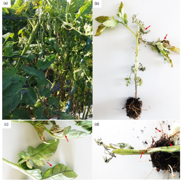
Symptomatology and pathogen isolation: Typical symptoms of Fusarium wilt were observed at all growth stages of tomato plants in Lam Dong. Diseased tomato plants typically showed yellowing and wilting of basal leaves, often beginning on one side of the plant or a single branch before spreading throughout the canopy. Leaves turned yellow and wilted but generally did not abscise (Fig. 1a-b). Characteristic symptoms included sudden partial wilting of leaves or leaflets, frequently unilateral, with veins remaining prominent (Fig. 1c). Green wilting was more common, although yellow wilting was also observed. In advanced cases, vascular tissues displayed partial discoloration, and severe infections led to plant death. The stem base of severely diseased plants often became shriveled, brown, and dry-rotted, accompanied by vascular discoloration. Similar symptoms were also evident in the roots of advanced infections (Fig. 1d).
|
Morphological characterization
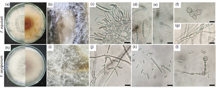
Fusarium incarnatum-equiseti species complex (FIESC): Seven isolates were classified within this complex, showing consistent morphological features (Fig. 2a-g). On PDA, colonies were white to slightly yellow or light brown, featuring sharp, irregular margins and abundant aerial hyphae (Fig. 2a). On CLA, all isolates produced sporodochia with an abundance of mostly macroconidia (Fig. 2b-c). These macroconidia were sharply curved with pointed basal and apical cells. They were typically produced as false heads on highly branched conidiophores, where sporodochial conidiophore cells appeared flask-shaped, being characteristically short and broad at the base (Fig. 2c-d). While microconidia were absent, mesoconidia were occasionally observed (Fig. 2e). Chlamydospores were round to obovoid, thick-walled, and formed singly, in pairs, or in chains and clusters (Fig. 2f-g). These traits closely match descriptions of members of the FIESC in FusaHelp (Comparative morphometric dimensions and key diagnostic features are summarized in Table 1.
|
| Table 1: | Morphological and morphometric characteristics of Fusarium incarnatum–equiseti (FIESC) and F. oxysporum (FOSC) species complexes | |||
| Characteristics | FIESC (7 isolates) | FOSC (Isolate To7031) |
| Sporodochia (on CLA) | Abundant, formed within 2-3 weeks | Absent, even after 30 days |
| Macroconidia shape | Sharply curved, pointed ends | Straight or slightly curved, foot-shaped base |
| Macroconidia length (μm)* | 28.48-56.10 (41.61±6.84) | 24.75-43.95 (33.59±3.56) |
| Macroconidia width (μm)* | 3.65-5.39 (4.32±0.39) | 3.12-4.91 (3.85±0.42) |
| Macroconidia septa | 03-Jun | 3-5 (mostly 3) |
| Microconidia | Absent | Present |
| Microconidia length (μm)* | N/A | 8.05-15.79 (11.33±2.30) |
| Microconidia width (μm)* | N/A | 2.37-5.08 (3.60±0.70) |
| Microconidia shape | N/A | Elongated ovoid, kidney-shaped (0-1 septate) |
| Phialides type | Highly branched conidiophores | Simple monophialides |
| Chlamydospores | Singly, in pairs, or in chains/clusters | Typically single, terminal or intercalary |
| *Data are presented as range (Mean±Standard Deviation) and n = 40 per spore type | ||
Fusarium oxysporum species complex (FOSC): One isolate was assigned to this group (Fig. 2h-l). On PDA, colonies appeared white and circular with abundant aerial hyphae (Fig. 2h). Notably, the isolate did not form sporodochia on CLA even after 30 days of incubation (Fig. 2i). The isolate produced three distinct spore types: microconidia, macroconidia, and chlamydospores. Microconidia were elongated ovoid, maggot- or kidney-shaped and were formed in false heads on short, simple monophialides a hallmark of this complex (Fig. 2j). Macroconidia were mostly straight or slightly curved, featuring a pointed basal cell and a characteristic foot-shaped apical cell (Fig. 2k). Chlamydospores were typically round, and formed either terminally or internally, and occurred mostly single (Fig. 2l). The detailed morphometric data for all spore types are presented in Table 1. Based on these features, isolate To7031 was confirmed as F. oxysporum.
|
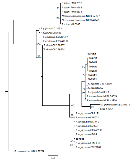
Molecular identification: Molecular identification of fungal isolates was carried out through a combination of BLAST searches against the NCBI database and phylogenetic analyses. The BLAST results corroborated the morphological identification. All isolates assigned to the FIESC group exhibited 99.6–100% sequence identity with reference accessions of the FIESC complex (e.g., MT560337.1, MT428185.1, MH054915.1), thereby confirming their affiliation with Fusarium equiseti. Likewise, the single FOSC isolate (To7031) displayed >99% sequence identity with multiple F. oxysporum accessions (e.g., KY910846.1).
|
Phylogenetic reconstruction further resolved the tomato isolates into two distinct clades, corresponding to FOSC and FIESC. Isolate To7031 clustered with the F. oxysporum reference strain KJH002 from GenBank, whereas all FIESC isolates grouped with F. equiseti reference sequences, supported by high bootstrap values (Fig. 3). Collectively, these results provide robust evidence that the isolates under investigation belong to F. oxysporum and F. equiseti, with the latter representing the more epidemiologically significant pathogen due to its higher frequency of isolation.
Pathogenicity tests
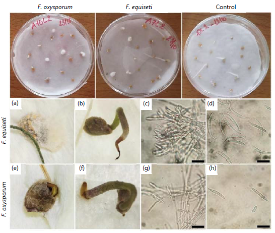
Blotter assay: In the blotter assay, seeds infected with Fusarium spp. developed a superficial mycelial layer, resulting in seed rot and failure to germinate (Fig. 4a-e). Post-emergent seedlings arising from infected seeds commonly exhibited disease symptoms, including root tip and collar rot as well as sprout necrosis (Fig. 4b-f), which ultimately led to seedling mortality. The fungal growth on seeds differed in appearance, with purple-white mycelia for F. oxysporum (Fig. 4a-d) and orange-white for F. equiseti (Fig. 4e-h); however, post-germination symptoms were indistinguishable between the two species (Fig. 4).
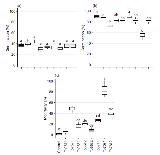
With respect to germination, Fusarium isolates exerted little influence at 5 DAI, when germination averaged ~40% across all treatments without significant differences. By 10 DAI, germination reached 80-90% in most treatments, including the control, but was significantly reduced in seeds inoculated with F. oxysporum To7031 (54.9%) and F. equiseti To2521 (72.5%) (p<0.05) (Fig. 5a-b).
|
Seedling mortality also varied significantly among isolates. While nearly all control seedlings remained healthy throughout the assay, mortality at 10 DAI was highest in F. oxysporum To7031 (83.3%), followed by F. equiseti To2521 (48.5%). Most other isolates induced <20% mortality, although F. equiseti To7812 and To6711 exceeded this level (Fig. 5c).
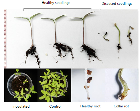
Pot assay: Results from the pot assay demonstrated that all Fusarium isolates were pathogenic to tomato, adversely affecting both seed germination and seedling development under seed-inoculated conditions. Infection by Fusarium spp. resulted in seed rot, reduced germination, and the development of collar rot in emerged seedlings (Fig. 6 and 7).
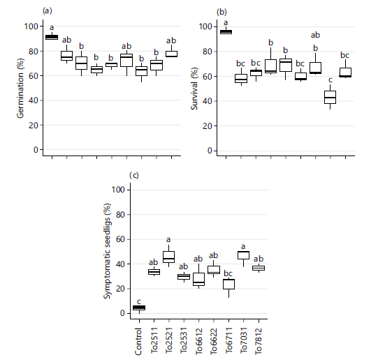
Germination percentage was highest in the control (>90%) and significantly reduced (<80%) across all inoculated treatments, although differences among isolates were minimal. By 8 DAS, most isolates significantly suppressed germination relative to the control, except for F. equiseti isolates To2511, To6622, and To7812, which did not differ significantly from either the control or other treatments (p>0.05) (Fig. 7a).
Seedling survival was also markedly reduced by infection. While nearly all germinated seeds in the control developed into healthy seedlings, only 43-70% of plants derived from inoculated seeds survived to 16 DAS, a level significantly lower than the control (p<0.05). Among the isolates, F. oxysporum To7031 caused the most severe reduction (43.2%) in post-emergence survival (Fig. 7b).
|
|
Infected seedlings also exhibited characteristic disease symptoms, including root rot, collar rot, hypocotyl browning, sprout necrosis, and abnormal growth (Fig. 6). Whereas >96% of control seedlings remained symptomless (only 3.7% abnormal growth), approximately 23-46% of infected seedlings displayed these symptoms, with the most severe effects recorded for F. oxysporum To7031 (45.8%) and F. equiseti To2521 (45.8%) (Fig. 7c).
Finally, representative diseased seedlings and non-germinated seeds were collected for microscopic examination and fungal re-isolation. Subsequent morphological identification confirmed the causal association of the isolates, thereby fulfilling Koch’s postulates.
To sum up, both blotter and pot assays confirmed the pathogenicity of all tested Fusarium isolates on tomato seeds and seedlings. In both assays, F. oxysporum To7031 consistently exhibited the strongest pathogenic effects, causing the greatest reductions in germination, and seedling survival, as well as the highest levels of mortality and symptomatic seedlings. Fusarium equiseti isolates, particularly To2521, also showed considerable pathogenicity, though with moderate variation among isolates. Overall, the results demonstrate that Fusarium seed infection leads to pre- and post-emergence seedling mortality, and disease symptoms, with clear differences in virulence between isolates.
DISCUSSION
Fusarium wilt remains one of the most destructive diseases of tomato worldwide, threatening production across virtually all growing regions3. Although F. oxysporum has long been considered the principal causal agent, increasing evidence points to the involvement of additional Fusarium species in tomato wilt epidemics, highlighting the complexity of this pathosystem12,13,26. In this study, tomato plants in Lam Dong Province, Vietnam, were found to exhibit classical wilt symptoms, and subsequent analyses confirmed that both F. oxysporum and F. equiseti were associated with the disease. This provides new evidence that tomato Fusarium wilt in Vietnam is not caused by a single species but by a pathogen complex.
Morphological observations supported by molecular characterization enabled the accurate identification of the isolates. While colony traits and conidial morphology allowed preliminary separation into the FOSC (F. oxysporum species complex) and FIESC (F. incarnatum–equiseti species complex), definitive identification was achieved through ITS sequence analysis and phylogeny. The morphological features observed in this study were highly consistent with previous descriptions27, underscoring the value of combining traditional and molecular approaches for identifying morphologically similar Fusarium taxa.
Pathogenicity assays confirmed that both species induced typical wilt symptoms, beginning with root tip and collar rot and progressing to vascular browning and plant death. These results are consistent with previous reports of F. equiseti causing seedling wilt in tomato28 and chickpea29. Importantly, the symptoms caused by the two pathogens were indistinguishable in seedlings, and both were recovered from field plants exhibiting identical wilt. This convergence complicates diagnosis and suggests that field-level assessments alone are insufficient to distinguish among causal species. Instead, molecular tools will be required for precise detection and monitoring.
The predominance of F. equiseti over F. oxysporum in Vietnam is particularly noteworthy. Unlike the situation in India, where F. oxysporum remains the dominant wilt pathogen26, or Indonesia, where F. equiseti occurs sporadically14, our findings align with Pakistan, where it accounted for 69% of tomato wilt cases13. Its frequent isolation from wilted tomato plants in Lam Dong indicates that F. equiseti already contributes substantially to local disease pressure and may become endemic, affecting tomato and other vegetable hosts. Although traditionally linked with root rot in various crops18,29,30, its increasing detection as a wilt pathogen highlights its emerging global significance. While F. oxysporum remains the most
widespread tomato wilt pathogen worldwide26, the dominance of F. equiseti in Lam Dong suggests a shifting epidemiological landscape, where F. oxysporum may drive severe outbreaks through higher aggressiveness, but F. equiseti poses the greater long-term threat due to its higher prevalence and persistence.
The discovery of F. equiseti as a predominant tomato wilt pathogen in Vietnam is particularly significant for several reasons. First, it indicates that the epidemiology of Fusarium wilt is more complex than previously assumed and involves a broader spectrum of pathogens. Second, the predominance of F. equiseti in Lam Dong, in contrast with the dominance of F. oxysporum in other countries17,26, raises the possibility that local agro-ecological conditions, cropping systems, or host genotypes may favor the establishment of this species. Finally, the finding underscores the need to reconsider current diagnostic and management strategies, which have historically focused on F. oxysporum alone.
An additional concern highlighted in this study is the seed-borne transmission of both pathogens. Infected seeds exhibited fungal colonization, impaired germination, and high seedling mortality. This confirms that Fusarium wilt pathogens are not only soil-borne31 but also seed-transmissible, which has important implications for disease spread and management. Seed-borne inoculum can establish disease in previously disease-free fields or after soil disinfestation, undermining control efforts32,33. Furthermore, the trade of infected seed may facilitate long-distance and even international dissemination of the pathogens34. Given the predominance of F. equiseti in this study, its seed-borne nature represents a substantial phytosanitary risk, emphasizing the urgent need for seed testing, certification, and strict quarantine regulations to limit further spread32.
Taken together, our findings demonstrate that tomato wilt in Vietnam is caused by at least two Fusarium species: The well-known cosmopolitan pathogen F. oxysporum and the increasingly reported F. equiseti. The predominance of the latter suggests that it is not only emerging globally but may already play a leading role in certain regions. The indistinguishable symptoms produced by the two species complicate diagnosis and management, and their ability to spread via seed represents a major risk to disease control and biosecurity. Future studies should focus on broader surveys across Vietnam to determine the distribution and diversity of F. equiseti in tomato and other hosts, as well as on comparative studies of their pathogenicity mechanisms and responses to management practices. Developing rapid molecular diagnostic tools and integrating them into seed health programs will be essential steps toward sustainable tomato production in Vietnam and beyond.
CONCLUSION
This study demonstrates that tomato Fusarium wilt in Lam Dong Province, Vietnam, is associated with a pathogen complex comprising Fusarium oxysporum and Fusarium equiseti. Morphological, molecular, and pathogenicity analyses confirmed that both species are capable of inducing typical wilt symptoms and significant seedling mortality under controlled conditions. Notably, F. equiseti was more frequently isolated, indicating its increasing dominance in the local disease complex and suggesting a potential epidemiological shift in the causal agents of tomato wilt. The inability to distinguish infections caused by these species based on symptoms alone highlights the limitations of field diagnosis and underscores the importance of molecular identification for accurate detection. Furthermore, evidence of seed-borne transmission emphasizes the risk of long-distance dissemination and the need for improved seed health management. Overall, the findings highlight the emergence of F. equiseti as a significant pathogen of tomato and reinforce the necessity for continuous surveillance, improved diagnostic tools, and integrated disease management strategies to protect tomato production in Vietnam.
SIGNIFICANCE STATEMENT
Tomato wilt disease is a serious problem that reduces crop yield and threatens farmer livelihoods in Vietnam, yet the exact causes are not always clearly understood. This study identifies the fungi responsible for this disease in Lam Dong, one of the country’s main tomato-growing regions, and evaluates how harmful they are to young plants. Samples were collected from diseased plants, and the fungi were identified using their physical characteristics and DNA analysis. It shows that most samples belonged to a species called Fusarium equiseti, while a smaller number were F. oxysporum. Both types were able to infect tomato seeds and seedlings, causing rot, poor germination, stem damage, and plant death. Importantly, the symptoms caused by these fungi looked the same, making it difficult for farmers to tell them apart in the field. The study reveals that F. equiseti, previously considered less important, is now a major cause of tomato wilt in this region. These findings will help improve disease diagnosis, guide better management practices, and support efforts to protect tomato production and farmer income.
ACKNOWLEDGMENTS
The author acknowledges the Ministry of Education and Training of Vietnam for supporting a previous project (2023), whose research contributed to this work, and Dalat University for funding the completion of most of the study. The author is also grateful to the Faculty of Agriculture and Forestry, Dalat University, for providing facilities, equipment, and technical support, and acknowledges Tran Thi Minh Loan, Cao Thi Lan, Pham Ngoc Tuan, and Dinh Quang Anh (Faculty of Agriculture and Forestry, Dalat University) for their assistance.
FUNDING
This work was supported by the Science and Technology Development Fund of Dalat University, Vietnam. The authors declare that no specific grant number is applicable for this institutional funding.
REFERENCES
- Mottet, A., C. de Haan, A. Falcucci, G. Tempio, C. Opio and P. Gerber, 2017. Livestock: On our plates or eating at our table? A new analysis of the feed/food debate. Global Food Secur., 14: 1-8.
- Liu, Z., J. Jiang, A. Ren, X. Xu and H. Zhang et al., 2021. Heterosis and combining ability analysis of fruit yield, early maturity, and quality in tomato. Agronomy, 11.
- Srinivas, C., D.N. Devi, K.N. Murthy, C.D. Mohan and T.R. Lakshmeesha et al., 2019. Fusarium oxysporum f. sp. lycopersici causal agent of vascular wilt disease of tomato: Biology to diversity-A review. Saudi J. Biol. Sci., 26: 1315-1324.
- Panno, S., S. Davino, A.G. Caruso, S. Bertacca and A. Crnogorac et al., 2021. A review of the most common and economically important diseases that undermine the cultivation of tomato crop in the Mediterranean basin. Agronomy, 11.
- Yan, X., S. Guo, K. Gao, S. Sun, C. Yin and Y. Tian, 2023. The impact of the soil survival of the pathogen of Fusarium wilt on soil nutrient cycling mediated by microorganisms. Microorganisms, 11.
- Catanzariti, A.M., G.T.T. Lim and D.A. Jones, 2015. The tomato I-3 gene: A novel gene for resistance to Fusarium wilt disease. New Phytol., 207: 106-118.
- Takken, F. and M. Rep, 2010. The arms race between tomato and Fusarium oxysporum. Mol. Plant Pathol., 11: 309-314.
- Rodríguez-Molina, M.C., M.B. García-García, M.D. Osuna, A. de Llano, M.M. Guerrero-Díaz and P. Serrano-Pérez, 2025. First report of Fusarium oxysporum f. sp. lycopersici race 3 on tomato in Spain. New Dis. Rep., 51.
- Li, J., L. Fokkens, L.J. Conneely and M. Rep, 2020. Partial pathogenicity chromosomes in Fusarium oxysporum are sufficient to cause disease and can be horizontally transferred. Environ. Microbiol., 22: 4985-5004.
- Adhikari, T.B., A. Gao, T. Ingram and F.J. Louws, 2020. Pathogenomics characterization of an emerging fungal pathogen, Fusarium oxysporum f. sp. lycopersici in greenhouse tomato production systems. Front. Microbiol., 11.
- Vawdrey, L.L. and R.A. Peterson, 1988. Fusarium solani, the cause of foot rot of tomatoes in Central Queensland. Australas. Plant Pathol., 17: 24-25.
- Nandan, M., S. Hiremath, H.D.V. Kumar, M. Mantesh, C.R.J. Basha and C.N.L. Reddy, 2020. Morphological and molecular characterization of Fusarium solani infecting tomato. Plant Dis. Res., 35: 111-117.
- Akbar, A., S. Hussain and G.S. Ali, 2018. Germplasm evaluation of tomato for resistance to the emerging wilt pathogen Fusarium equiseti. J. Agric. Stud., 6: 174-189.
- Darmadi, A.A.K., N.L. Suriani, S.K. Sudirga and K. Khalimi, 2019. First study on Fusarium equiseti: Causes fusarium wilt in tomato crop in Bali, Indonesia. Sabrao J. Breed. Genet., 51: 442-450.
- Severo, R., L.J.S. Shibutani, G.F. da Silva, S. da Silva Costa Guimarães and F. de Alcântara Neto et al., 2024. Fusarium species causing root rot and wilt in tomato in Brazil. J. Phytopathol., 172.
- Deng, S., Q. Liu, W. Chang, J. Liu and H. Wang, 2023. First specific detection and validation of tomato wilt caused by Fusarium brachygibbosum using a PCR assay. PeerJ, 11.
- Isaac, M.R., S.G. Leyva-Mir, J. Sahagún-Castellanos, K. Câmara-Correia, J.M. Tovar-Pedraza and J.E. Rodríguez-Pérez, 2018. Occurrence, identification, and pathogenicity of Fusarium spp. associated with tomato wilt in Mexico. Notulae Botanicae Horti Agrobotanici Cluj-Napoca, 46: 484-493.
- Beltran, M., J.C. Delgado, A.G. Valdivia, A. Hernandez and A.M. Garcia, 2023. First report of Fusarium equiseti causing root and crown rot in tomato in Mexico. Plant Dis., 107.
- Ávalos-Sánchez, E., A. López-Martínez, F.D. Molina-Aiz, J. Reca, P. Marín-Membrive and D.L. Valera-Martínez, 2022. Effect of different substrates, and irrigation with water with different saline concentrations, on the development of tomato fungal diseases in an Almería-type greenhouse. Agronomy, 12.
- Seidel, D., S. Wurster, J.D. Jenks, H. Sati and J.P. Gangneux et al., 2024. Impact of climate change and natural disasters on fungal infections. Lancet Microbe, 5: e594-e605.
- Sahabne Ullah, D. Mostert, K. Serfontein and A. Viljoen, 2021. The survival and treatment of Fusarium oxysporum f. sp. cubense in water. J. Fungi, 7.
- Vũ, N.T., H. Quyền, D.H. Xô and L.Q. Luân, 2017. Preparation of the oligochitosan-silver nanoparticles with a potential application as an antigungal fertilizer against Fusarium oxysporum causing fusarium wilt disease on tomato. Vietnam J. Biotechnol., 15: 141-149.
- Nguyen, P.V., H.V.L. Nguyễn, Đ.T. Lê and H.H.T. Nguyễn, 2024. Enhancement of tomato growth and biocontrol of Fusarium solani root rot disease by Streptomyces rochei BT02. BIOTROPIA, 31: 106-113.
- Le, D., 2024. Unveiling Fusaria mycoflora associated with natural occurrence of lisianthus wilt and stem rot in central-highland Vietnam. Arch. Microbiol., 206.
- Ihaka, R. and R. Gentleman, 1996. R: A language for data analysis and graphics. J. Comput. Graphical Stat., 5: 299-314.
- Pothiraj, G., Z. Hussain, A.K. Singh, A.U. Solanke, R. Aggarwal, R. Ramesh and V. Shanmugam, 2021. Characterization of Fusarium spp. inciting vascular wilt of tomato and its management by a Chaetomium-based biocontrol consortium. Front. Plant Sci., 12.
- Leslie, J.F. and B.A. Summerell, 2006. The Fusarium Laboratory Manual. Blackwell Publishing, United States, ISBN: 9780813819198, Pages: 388.
- Bacon, C.W., I.E. Yates, D.M. Hinton and F. Meredith, 2001. Biological control of Fusarium moniliforme in maize. Environ. Health Perspect., 109: 325-332.
- El Hazzat, N., M. Adnani, S. Msairi, M.A. El Alaoui and N. Mouden et al., 2023. Fusarium equiseti as one of the main Fusarium species causing wilt and root rot of chickpeas in Morocco. Acta Mycol., 57.
- Yezli, W., N. Hamini-Kadar, N. Zebboudj, L. Blondin, D. Tharreau and M. Kihal, 2019. First report of crown and root rot of tomato caused by Fusarium equiseti in Algeria. J. Plant Pathol.
- Khurshid, S., A. Shoaib, A. Javaid, F. Akhtar, M. Shafiq and U. Qaisar, 2017. Management of Fusarium wilt of tomato by soil amendment with Cenchrus pennisetiformis under chromium stress. Physiol. Mol. Plant Pathol., 97: 58-68.
- Vishunavat, K., K. Prabakar and T. Anand, 2023. Seed Health: Testing and Management. In: Seed Science and Technology: Biology, Production, Quality, Dadlani, M. and D.K. Yadava (Eds.), Springer Nature, Singapore, ISBN: 978-981-19-5888-5, pp: 335-364.
- Moumni, M., G. Brodal and G. Romanazzi, 2023. Recent innovative seed treatment methods in the management of seedborne pathogens. Food Secur., 15: 1365-1382.
- Martín, I., L. Gálvez, L. Guasch and D. Palmero, 2022. Fungal pathogens and seed storage in the dry state. Plants, 11.
How to Cite this paper?
APA-7 Style
Le,
D. (2026). Emerging Role of Fusarium equiseti in Tomato Fusarium Wilt in Vietnam. Asian Journal of Plant Pathology, 20(1), 18-32. https://doi.org/10.3923/ajpp.2026.18.32
ACS Style
Le,
D. Emerging Role of Fusarium equiseti in Tomato Fusarium Wilt in Vietnam. Asian J. Plant Pathol. 2026, 20, 18-32. https://doi.org/10.3923/ajpp.2026.18.32
AMA Style
Le
D. Emerging Role of Fusarium equiseti in Tomato Fusarium Wilt in Vietnam. Asian Journal of Plant Pathology. 2026; 20(1): 18-32. https://doi.org/10.3923/ajpp.2026.18.32
Chicago/Turabian Style
Le, Dung.
2026. "Emerging Role of Fusarium equiseti in Tomato Fusarium Wilt in Vietnam" Asian Journal of Plant Pathology 20, no. 1: 18-32. https://doi.org/10.3923/ajpp.2026.18.32

This work is licensed under a Creative Commons Attribution 4.0 International License.